Home
/ Pelvic Anatomy Female : Female Pelvic Anatomy Artwork Stock Image C010 7098 Science Photo Library : Use the mouse scroll wheel to move the images up and down alternatively use the tiny arrows (>>) on both side of the image to move the images.>>) on both side of the image to move the images.
Pelvic Anatomy Female : Female Pelvic Anatomy Artwork Stock Image C010 7098 Science Photo Library : Use the mouse scroll wheel to move the images up and down alternatively use the tiny arrows (>>) on both side of the image to move the images.>>) on both side of the image to move the images.
Pelvic Anatomy Female : Female Pelvic Anatomy Artwork Stock Image C010 7098 Science Photo Library : Use the mouse scroll wheel to move the images up and down alternatively use the tiny arrows (>>) on both side of the image to move the images.>>) on both side of the image to move the images.. The pelvis's frame is made up of the bones of the pelvis, which connect the axial skeleton to the femurs, and therefore acts in weight bearing of the upper body. Magnetic resonance imaging or mri of the female pelvis offers a unique display of the pelvic anatomy, including a woman's ovaries, uterus, and fallopian tubes. This mri female pelvis sagittal cross sectional anatomy title tool is absolutely free to use. Imaging case of the week 69. Each pelvic bone (hip bone) is made by the combination three bones namely, the ilium, pubis, and ischium.
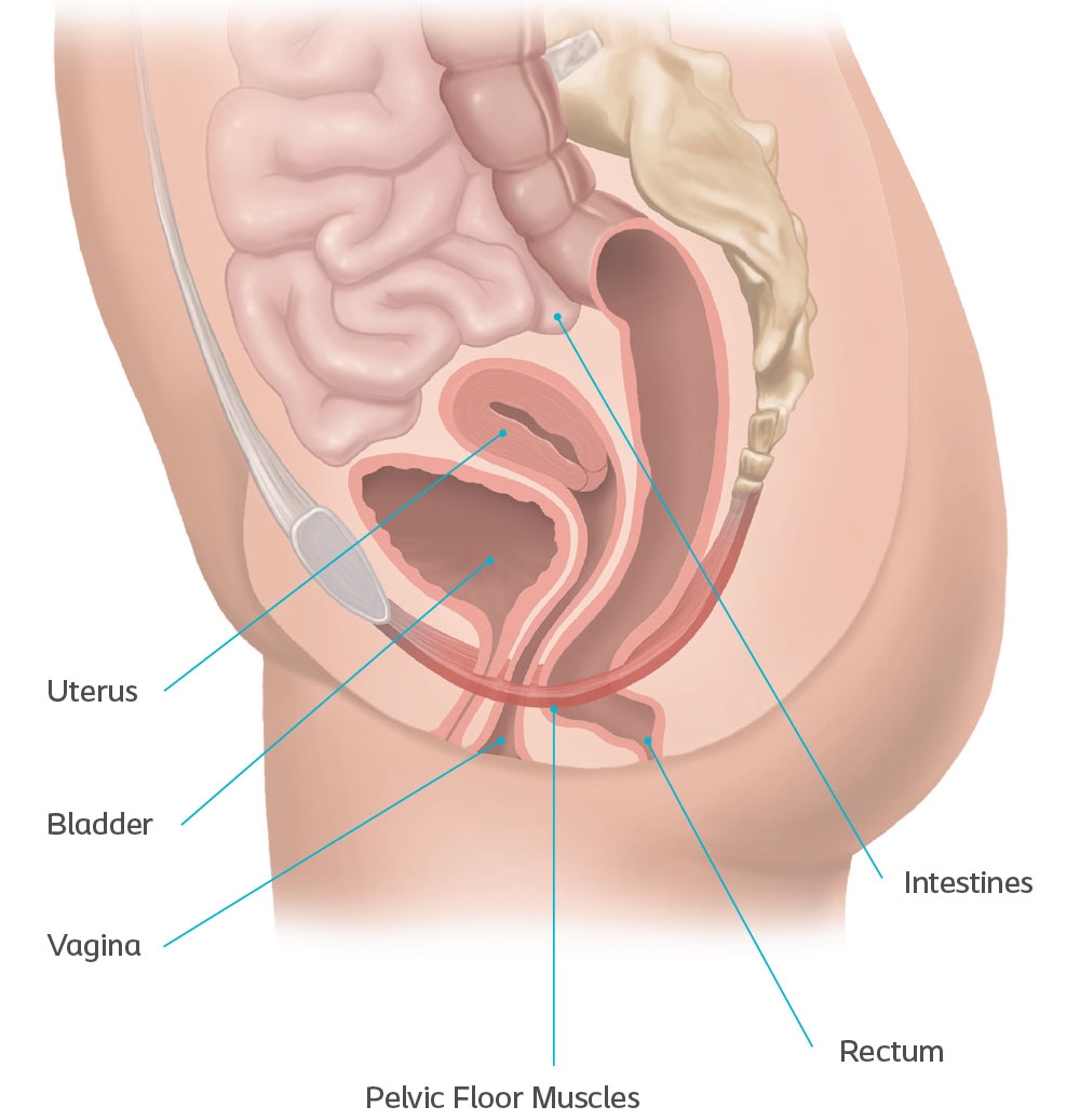
In women, the pelvis houses the uterus, tubes, ovaries and vagina. Hi and welcome to my functional female pelvic anatomy course! Magnetic resonance imaging or mri of the female pelvis offers a unique display of the pelvic anatomy, including a woman's ovaries, uterus, and fallopian tubes. Pelvic diaphragm —the term pelvic diaphragm refers to the levator ani muscle and its covering fasciae, both the superior fascia and the inferior fascia. The pelvis is the lower part of the torso.
Learn More About Pelvic Organ Prolapse Symptoms Treatments from www.femalepelvicsolutions.com Enroll in course for €97. One major difference between males and females is their. A comprehensive overview of functional anatomy & assessment of the female pelvis. Describe the anatomy of the pelvic wall, bones, joints & muscles. 1.1 superficial perineal compartment (superficial transverse perineal muscles ( a ), vestibular and bartholin's gland after left bulbospongiosus muscle removal ( b ) (with kind permission from springer science + business media: Concept for study of anatom. Magnetic resonance imaging or mri of the female pelvis offers a unique display of the pelvic anatomy, including a woman's ovaries, uterus, and fallopian tubes. Its primary function is the transmission of forces fro.
Describe the anatomy of the pelvic wall, bones, joints & muscles.
Enroll in course for €97. Anatomy of the pelvic floor. Surgical anatomy of the female pelvis by laparoscopy. Anatomy xray of the shoulder joint. It's located between the abdomen and the legs. 1 chapter 1 applied clinical obstetric anatomy nasrat1949. Differentiate the different types of the female pelvis. The term pelvic floor refers to all of the supportive structures that are involved with pelvic organ support. Each pelvic bone (hip bone) is made by the combination three bones namely, the ilium, pubis, and ischium. Its primary function is the transmission of forces fro. Pelvic diaphragm —the term pelvic diaphragm refers to the levator ani muscle and its covering fasciae, both the superior fascia and the inferior fascia. They act to support the female viscera and provide a conduit for neurovascular structures. Explore more like female pelvic anatomy ligaments.
Its primary function is the transmission of forces fro. Use the mouse scroll wheel to move the images up and down alternatively use the tiny arrows (>>) on both side of the image to move the images.>>) on both side of the image to move the images. The obstetrical anatomy of a typical female pelvis is best considered as one unit. Differentiate the different types of the female pelvis. Explore more like female pelvic anatomy ligaments.
Https Www Hopkinsmedicine Org Gynecology Obstetrics Pdfs Residency Anatomy Pelvicanatomyforresident Pdf from By continuing to browse this site you are agreeing to our use of cookies. Thakar and fenner 5 , figure 1.2, p. 1 chapter 1 applied clinical obstetric anatomy nasrat1949. It's located between the abdomen and the legs. 3.9 out of 5 stars. On a sagittal plane, the uterus has a pyriform shape: The obstetrical anatomy of a typical female pelvis is best considered as one unit. {{configctrl2.info.metadescription}} this site uses cookies.
The pelvis is the lower portion of the trunk, located between the abdomen and the lower limbs.
The uterus represents the essential landmark of pelvic anatomy. A comprehensive overview of functional anatomy & assessment of the female pelvis. Describe the boundaries and subdivisions of the pelvis. One major difference between males and females is their. In this image, you will find anatomy of female pelvic area in detail, suspensory ligament of ovary, paravesical pouch, broad ligament, mesovarium, ovary, uterine (fallopian) tube, round ligament of uterus, ligament of ovary, uterus, internal iliac artery and vein, linea terminalis, cervix, obturator membrane, obturator fascia in it. On a sagittal plane, the uterus has a pyriform shape: Ct body (lymph nodes) ct. Female anatomy includes the external genitals, or the vulva, and the internal reproductive organs, which include the ovaries and the uterus. This mri female pelvis sagittal cross sectional anatomy title tool is absolutely free to use. Gynecoid/ genuine pelvis, the brim is round, more wider, and both ischial spines are less prominent this allows easy baby delivery. Each pelvic bone (hip bone) is made by the combination three bones namely, the ilium, pubis, and ischium. Laparoscopic anatomy of the female pelvic region. The superior two thirds correspond to the uterine body and the inferior third to the cervix.
Describe the anatomy of the pelvic wall, bones, joints & muscles. List the arterial & nerve supply list the lymph & venous drainage of the pelvis. Laparoscopic anatomy of the female pelvic region. This mri female pelvis sagittal cross sectional anatomy title tool is absolutely free to use. {{configctrl2.info.metadescription}} this site uses cookies.
5 Anatomical Detail Of Female Pelvic Anatomy Adapted From Netter 2002 Download Scientific Diagram from www.researchgate.net Female anatomy includes the external genitals, or the vulva, and the internal reproductive organs, which include the ovaries and the uterus. The term pelvic floor refers to all of the supportive structures that are involved with pelvic organ support. Gynecoid/ genuine pelvis, the brim is round, more wider, and both ischial spines are less prominent this allows easy baby delivery. Thakar and fenner 5 , figure 1.2, p. Anatomy xray of the shoulder joint. Laparoscopic anatomy of the female pelvic region. On a sagittal plane, the uterus has a pyriform shape: In this image, you will find anatomy of female pelvic area in detail, suspensory ligament of ovary, paravesical pouch, broad ligament, mesovarium, ovary, uterine (fallopian) tube, round ligament of uterus, ligament of ovary, uterus, internal iliac artery and vein, linea terminalis, cervix, obturator membrane, obturator fascia in it.
Differentiate the different types of the female pelvis.
Gynecoid, anthropoid, android, and platypelloid. Describe the anatomy of the pelvic wall, bones, joints & muscles. On a sagittal plane, the uterus has a pyriform shape: 1.1 superficial perineal compartment (superficial transverse perineal muscles ( a ), vestibular and bartholin's gland after left bulbospongiosus muscle removal ( b ) (with kind permission from springer science + business media: This area provides support for the intestines and also contains the bladder and reproductive organs. A comprehensive & practical guide to female pelvic anatomy! Each pelvic bone (hip bone) is made by the combination three bones namely, the ilium, pubis, and ischium. Magnetic resonance imaging or mri of the female pelvis offers a unique display of the pelvic anatomy, including a woman's ovaries, uterus, and fallopian tubes. A comprehensive overview of functional anatomy & assessment of the female pelvis. Use the mouse scroll wheel to move the images up and down alternatively use the tiny arrows (>>) on both side of the image to move the images.>>) on both side of the image to move the images. Female anatomy includes the external genitals, or the vulva, and the internal reproductive organs, which include the ovaries and the uterus. Laparoscopic anatomy of the female pelvic region. Explore more like female pelvic anatomy ligaments.
The pelvis is the lower portion of the trunk, located between the abdomen and the lower limbs pelvic anatomy. Johns hopkins medicine, based in baltimore, maryland